Specialized Diagnostic Imaging Examinations
Symptoms of osteoarthritis may arise before the damage can be seen in standard X-rays. For this reason, radiologists at Hospital for Special Surgery often use the more sensitive , and forms of imaging, which are superior for detecting early osteoarthritis.
- MRI is very sensitive imaging that can reveal subtle changes in bony and soft tissues. An MRI can show a reactive bone edema , inflammation of soft tissues, as well as degenerated cartilage or bone fragments lodged in the joint. HSS uses a protocol of specific MRI pulse sequences to identify early evidence of cartilage degeneration. When evidence of cartilage wear is detected early, treatment can begin to prevent or delay progression. This in turn can postpone or eliminate the need for surgery.MRI of the hips showing osteoarthritis and edema of the femoral head and acetabulum.
- CT examinations, also called CT scans, are excellent for showing osteophytes and the ways they affect adjacent soft tissues. CT examinations are also useful in providing guidance for therapeutic and diagnostic procedures.CT scan of an arthritic showing hip bony femoral head debris
- Ultrasound is extremely sensitive for identifying synovial cysts that sometimes form in people with osteoarthritis. Ultrasound is excellent for evaluating the ligaments and tendons around the joint, which can be stretched or torn because of osteoarthritis.
What Are The Advantages Of Ultrasound Imaging In Rheumatoid Arthritis Diagnosis
A traditional method of monitoring the joint disease of patients with rheumatoid arthritis is X-rays, whereby images are produced by exposing photographic film . This technique has proven useful for doctors to follow the course of joint destruction. The early development of discrete bony destruction is associated with more severe rheumatoid disease. While standard X-ray radiographs contribute substantially to the clinical evaluation of rheumatoid arthritis, they do lack some sensitivity early in the course of disease. This means that substantial joint destruction must happen before changes on the standard X-ray test become apparent.
Modern treatment for rheumatoid arthritis is frequently directed at early disease. Accordingly, there efforts to establish methods for early diagnosis of the disease have increased. Several radiographic imaging modalities have been explored, including magnetic resonance imaging and ultrasonography. MRI scanning has been found to be sensitive as an indicator of early rheumatoid joint destruction, but it is very expensive and not widely available. Ultrasonography is an attractive method of imaging because of its low cost, absence of harmful radiation, and rapidity of imaging. Recent advances in ultrasound image technology have allowed the development of sonographic equipment for imaging inflamed joints in patients with rheumatoid arthritis.
Use Of Magnetic Resonance Imaging In Ra
Magnetic resonance imaging is a more sensitive imaging technique than an x-ray. An MRI is more useful in detecting changes to joints and tissues that resulted from inflammation. MRIs also detect joint damage earlier than traditional x-rays. One study showed that an MRI revealed more bony erosions in the hand than an x-ray.
In addition, MRIs are useful in detecting the thickening of synovial tissue that occurs with RA and swelling of bone marrow that predicts later bone erosion.1,3
Recommended Reading: What Is The Rheumatoid Arthritis Blood Test
Alternative Medicine For Arthritis
A variety of alternative therapies is used for arthritis. However, none of these has been approved by the FDA for the treatment of arthritis, so they may not be effective or safe. It is important to let your doctor know if you’re considering these types of treatments.
While some studies suggest that glucosamine and chondroitin supplements are as effective as NSAIDs for reducing pain, swelling, and stiffness in osteoarthritis, recent large studies funded by the NIH suggest these supplements are not very helpful, except perhaps in some cases. Typical daily doses are 1,500 milligrams for glucosamine and 1,200 milligrams for chondroitin.
The antibiotic doxycycline may have some potential to delay the progression of osteoarthritis by inhibiting enzymes that break down cartilage. More research is needed to confirm these results.
The NIH considers acupuncture an acceptable alternative treatment for osteoarthritis, especially if it affects the knee. Studies have shown that acupuncture helps reduce pain, may significantly lessen the need for painkillers, and can help increase range of motion in affected knee joints.
The supplement SAMe has been shown in some studies to be as effective for osteoarthritis pain as NSAIDs.
How Does An Mri Work

MRI creates a powerful magnetic field and radio waves to manipulate the position of hydrogen protons within your body. As the protons change position, they give off signals that can be picked up by the MRI scanner. These signals can be used by a computer to make an image of any tissue that is being scanned. Different tissues in the body contain various amounts of water and therefore more hydrogen. As a result, MRI images allow you to see the differences in these tissues.4
Recommended Reading: How Do You Know You Have Arthritis
Etiology And Risk Factors
Although osteoarthritis is especially common in older adults, its pathology of asymmetric joint cartilage loss, subchondral sclerosis , marginal osteophytes and subchondral cysts is the same in younger and older adults.1 Primary osteoarthritis is the most common form and is usually seen in weight-bearing joints that have undergone abnormal stresses .1316 The precise etiology of osteoarthritis is unknown, but biochemical and biomechanical factors are likely to be important in the etiology and pathogenesis.1 Biomechanical factors associated with osteoarthritis include obesity, muscle weakness and neurologic dysfunction. In primary osteoarthritis, the common sites of involvement include the hands, hips, knees and feet13,17. Secondary osteoarthritis is a complication of other arthropathies or secondary to trauma. Gout, rheumatoid arthritis and calcium pyrophosphate deposition disease are correlated with the onset of secondary osteoarthritis.
Home Remedies For Arthritis
In addition to treatments recommended by your doctor, you can use dry heat from a heating pad or moist heat in the form of a hot bath or a hot-water bottle wrapped in a towel to help relieve pain and stiffness. Heat and rest are very effective in the short run for most people with the disease. Regular exercise is also important to keep the joints mobile.
If you are overweight, losing weight is key, especially when arthritis affects the lower back, knees, and legs. Extra pounds add to the load and pressure on your joints, which can cause your arthritis to get worse faster. Being overweight also raises your chances of related health problems. Consult a registered dietitian who can help you plan a healthy weight loss program.
People with weakened, badly deformed fingers from rheumatoid arthritis benefit from specially designed utensils and door and drawer handles people with weakness in the legs and arms can use special bathroom fixtures, especially tub rails and elevated toilet seats.
Although arthritis may not be preventable, disability is — with a well-designed treatment program, including medications, exercise, and physical therapy when needed.
Here are some more things you can do to help keep the condition in check:
Educate yourself. Take a self-management course to learn specifics on day-to-day arthritis care.
Get active. Exercise can help you move better, lessen pain, and put off disability.
Don’t Miss: Reduce Arthritis Swelling In Fingers
Osteoarthritis: Signs And Symptoms
OA is diagnosed by a triad of typical symptoms, physical findings and radiographic changes. The American College of Rheumatology has set forth classification criteria to aid in the identification of patients with symptomatic OA that include, but do not rely solely on, radiographic findings.
Ra Imaging Tests: What Tests Are Done For Rheumatoid Arthritis
Rheumatoid arthritis imaging tests are used to look for signs of RA and to monitor the diseases progression. These tests primarily look for bone damage in the patients joints caused by the inflammation associated with RA.
X-rays used to be the most common form of imaging ordered, but they werent always great for reaching an early diagnosis. Todays more modern technology provides advanced imaging techniques like MRIs and ultrasounds, which allow doctors to find early signs of RA more easily.
All types of imaging tests are a critical component of diagnosing RA and monitoring the patients disease as it develops over time. Imaging tests provide doctors with a literal picture of the patients progression so that they can pursue appropriate treatment options.
You May Like: How To Ease Arthritis Pain In Fingers
What Does Arthritis Look Like On X
Arthritis is typically diagnosed on x-rays. Osteoarthritis is the most common form of arthritis and is related to wear-and-tear processes, genetics, injuries, and it is a normal part of the aging process. An arthritis joint will demonstrate narrowing of the space between the bones as the cartilage thins, bone spurs on the edges of the joint, small cysts within the bone, and sometimes deformity of the joint, causing it to look crooked. See the x-ray for common findings in osteoarthritis of the hand. The joints closest to the fingertip and the joint at the base of the thumb are the most common joints in the hand affected by osteoarthritis.
X-ray findings in OA of hand:
- joint space narrowing
- angular deformity or crooked finger
- subchondral cysts
Imaging And Nerve Tests For Arthritis
Imaging and nerve tests allow a doctor to see the internal structures without doing a medical procedure. So, these tests are commonly used both in the diagnosis as well as the monitoring of arthritis. These tests look for abnormalities of joints, organs, nerves and other signs of disease. These tests may also be necessary to help eliminate other causes of symptoms.
Imaging Tests
Also Check: Is Peanut Butter Bad For Arthritis
What Other Tests Are Used To Diagnose Psoriatic Arthritis
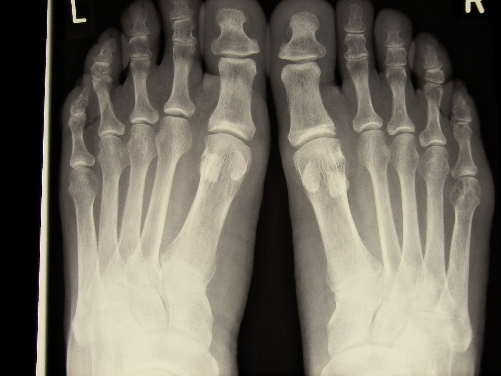
While X-rays are important to help determine damage by arthritis, such imaging tests cant confirm PsA alone. Part of this is due to the fact that other types of arthritis, such as rheumatoid arthritis , may look similar on X-rays.
To distinguish PsA from other autoimmune conditions that affect the joints, your doctor will need to run other exams and tests to provide an accurate diagnosis. These include:
How Arthritis In The Back Is Treated

Treatment for back arthritis depends on many factors, including your age, level of pain, type and severity of arthritis, other medical conditions and medications, and personal health goals. Because joint damage caused by arthritis is irreversible, treatment usually focuses on managing pain and preventing further damage.
Also Check: What Causes Arthritis Flare Up In Hands
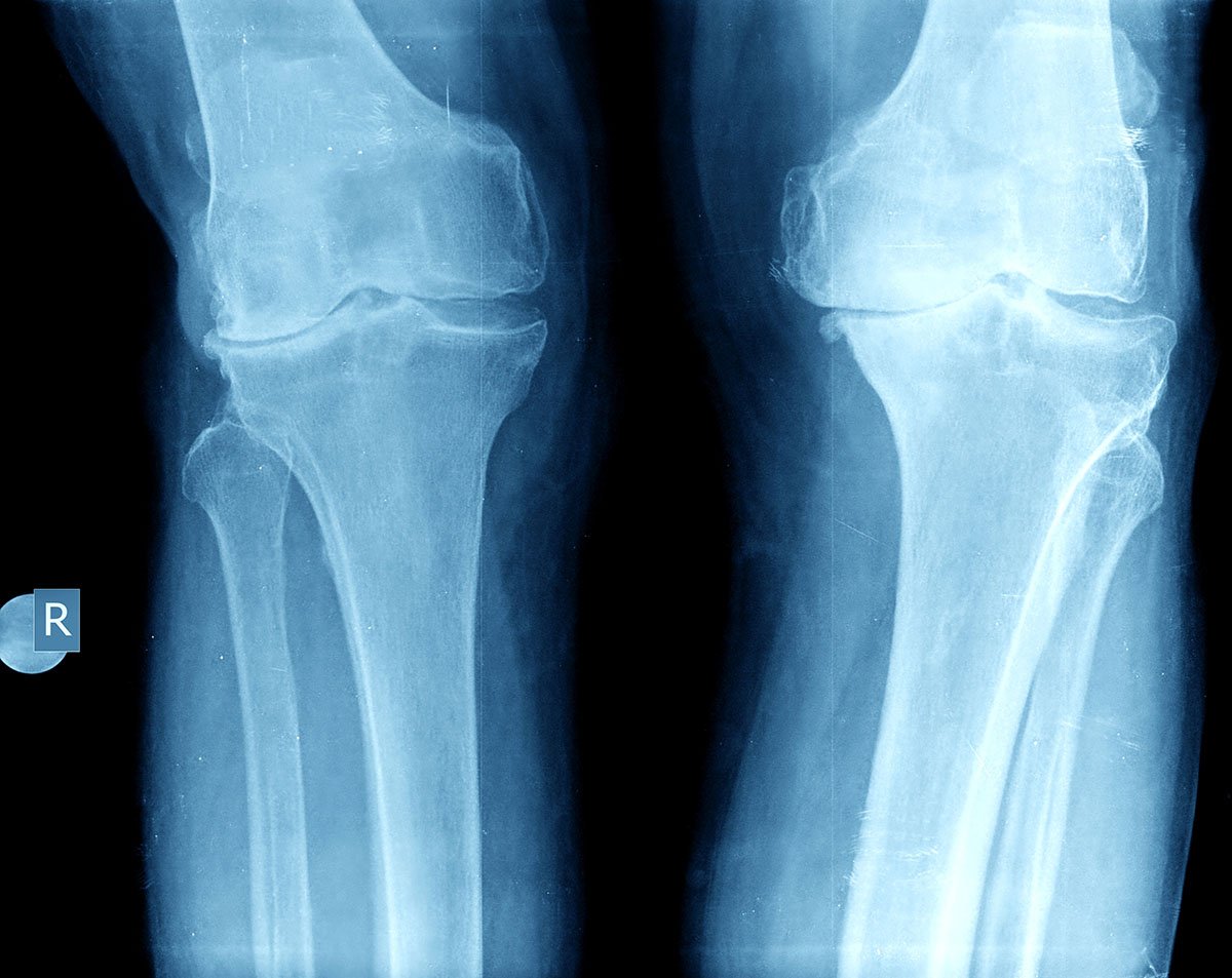
Signs Of Osteoarthritis In A Knee X
X-ray imaging results are usually available immediately after the procedure for you and your doctor to view. In some cases, your doctor may refer you to a specialist, such as a rheumatologist who specializes in arthritis, for further examination of your X-rays. This can take anywhere from a few days to a few weeks depending on your healthcare plan and the availability of the specialist.
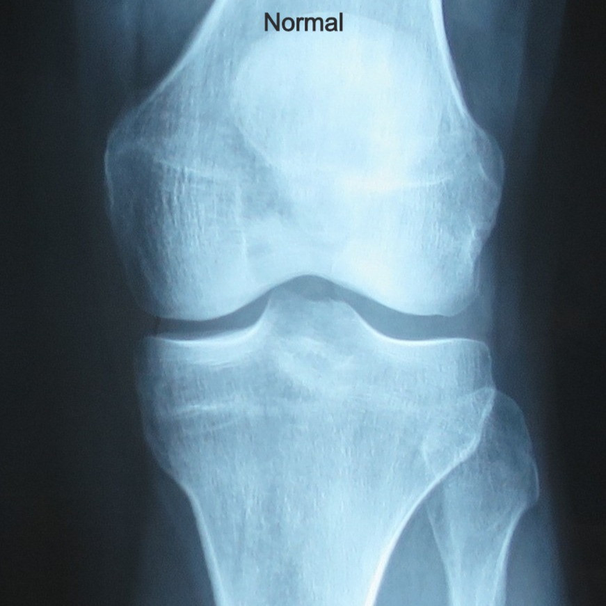
To check for osteoarthritis in your knee, your doctor will examine the bones of your knee joint in the image for any damage. Theyll also check the areas around your knee joints cartilage for any joint space narrowing, or cartilage loss in your knee joint. Cartilage isnt visible on an X-ray image, but joint space narrowing is the most obvious symptom of osteoarthritis and other joint conditions in which cartilage has eroded. The less cartilage thats left on your bone, the more severe your case of osteoarthritis.
Your doctor will also check for other signs of osteoarthritis, including osteophytes more commonly known as bone spurs. Bone spurs are growths of bone that stick out of the joint and can grind against each other, causing pain when you move your knee. Pieces of cartilage or bone can also break from the joint and get stuck in the joint area. This can make moving the joint even more painful.
Medical Imaging For Arthritis Diagnosis
Whether its magnetic resonance imaging , an ultrasound or a good old-fashioned X-ray, your doctor is likely to order some type of medical imaging to see whats going on below the surface with your arthritis.
The most important thing rheumatologists can do to assess patients is still a good history and clinical exam. The role of imaging is to assist in assessing the degree of severity, says Orrin Troum, MD, professor of medicine at University of Southern California and spokesperson for the International Society for Musculoskeletal Imaging in Rheumatology. Understanding its severity helps a doctor decide how aggressively to treat the disease.
You May Like: Can You Stop Arthritis In Fingers
Is Ultrasound Better Than X
In a study published in Arthritis& Rheumatism, ultrasound imaging was compared with standard X-ray imaging and shown to be superior at detecting bone erosions early in the course of rheumatoid arthritis.
In this study, 100 patients with rheumatoid arthritis underwent ultrasound and X-ray imaging of their hands. Twenty control patients were included in the ultrasound analysis for comparison. In the group of 100 RA patients, 127 abnormalities were detected in 56 patients by ultrasound, compared with 32 abnormalities in 17 patients detected by X-ray analysis. When patients with early rheumatoid arthritis were analyzed, 6.5-fold more abnormalities were detected by ultrasonography than by X-ray films. There were erosions detected by X-ray that were missed by ultrasound the correlation between erosions seen by X-ray and those seen by ultrasound was 86%.
From these results, the authors conclude that ultrasound is a reliable technique with greater sensitivity than standard X-ray radiography. They note that the ultrasound technique is influenced and limited by technical performance, requiring appropriate use of the sonographic equipment. Other limitations of this study include the technique used for analysis and patient selection .
What Causes Osteoarthritis
The most common form of arthritis, osteoarthritis, is associated with injuries, wear-and-tear processes, and genetics. An arthritis joint will demonstrate the narrow bone spaces due to various reasons. The cartilage thins, the formation of cysts within bones, bones spurs seen on the edges, deformity of joints are some of the reasons, which leads to crooked joints.
You can get rid of joint pain instantly by browsing through the best joint supplement reviews on the market. Read them carefully to arrive at the decision of buying the best product on the market.
You May Like: How To Deal With Arthritis
A Thorough Medical History
Your personal medical history is an important factor to consider when diagnosing PsA. Your doctor will ask questions about your symptoms, including their severity and when you first noticed them.
Additionally, your doctor will ask about any personal or family history of psoriasis, PsA, and other autoimmune conditions. Psoriasis may increase your chances of developing PsA, and both conditions can run in families.
Having a family history of autoimmune diseases may also increase your personal risk of developing PsA even if your parents or relatives have other types of autoimmune conditions.
Can You Tell If You Have Arthritis From An X
The image above is the X-Ray image of a patient that is diagnosed with osteoarthritis of the hand. Many things can be observed by looking at this image. In the case of osteoarthritis on the hand, the base of the thumb and joints near to the fingertips are the most commonly affected joints.
The image from the X-Ray clearly found these things:
- Joint Sclerosis
- Crooked Fingers
Don’t Miss: What Does The Rash From Psoriatic Arthritis Look Like
Needle Aspiration Of Joint Fluid
This is the only sure shot diagnostic method to confirm the diagnosis of gout. In the joints, there is the synovial fluid inside the synovial sac, which acts as a lubricant to facilitate joint movements.
This joint fluid is aspirated using a fine needle and syringe and tested for the presence of monosodium urate crystals . Sometimes in an acutely swollen and painful condition, this test may not be possible.
Summary of gout diagnosis
Diagnosis of gout can be confirmed as follows.
- Serum uric acid levels are high.
- Urine uric acid levels are not within the normal range. They may be either raised or lowered as explained above.
- X ray shows crystal deposits and/or damaged bones and cartilage.
- Synovial fluid shows the presence of uric acid crystals.
- Symptoms show that only one joint is affected.
- Symptoms and signs of gout are similar to the ones explained in gout symptoms.
How Is Arthritis Diagnosed And Evaluated

When diagnosing arthritis, your doctor will likely do a complete physical examination of your entire body, including your spine, joints, skin and eyes. You may undergo blood tests to detect markers of inflammation. In cases where an infection or gout is suspected, it may be useful to draw some fluid from a joint with a needle in order to analyze the contents of the material. In addition, your physician may order one or more of the following imaging tests:
Recommended Reading: How Can You Stop Arthritis In Your Hands
What Happens While Conducting An X
All the X-Ray tests are the part of a radiology department. The beam will be sent by an X-Ray machine for ionizing radiation via an X-Ray tube. The energy produced by the machine will pass through the body part that is being X-rayed.
After the energy is passed through the body part, the part of the body is captured on a digital camera or a film for the creation of a picture. The bones along with various other dense areas will be showed up as lighter shades of grey to white. There are some areas, which do not absorb the radiation. These areas will appear as dark grey to black color.
The X-Ray will not take much time. The entire test will be completed within 15 minutes, and there will be no discomfort during the test.